Critical Lower Limb Ischaemia
A 67 year-old male with history of type II diabetes mellitus, diabetic neuropathy, dyslipidemia, bilateral lower limb arterial occlusive disease and hypertension presented with bilateral lower extremity pain, more severe on the left side. The patient’s renal function was moderately reduced with an estimated glomerular filtration rate of 36 mL/min/m 2 . A lower extremity MRA study was requested to assess the extent of peripheral vascular disease and guide a treatment plan.
Technique:
Initial non-contrast MR angiography was performed with quiescent interval single shot (QISS) imaging, employing a tracking saturation pulse positioned towards the feet to suppress venous inflow, a non-selective saturation pulse to reduce signal from stationary tissues, and a fat-suppression pulse to further reduce signal from fat. QISS MRA is ECG gated, with diastolic-phase triggered data acquisition. Images were acquired from the feet upwards to the upper abdominal aorta.
Contrast-enhanced MR angiography was then performed with time-resolved MR angiography of the calves (TWIST) followed by bolus-chase MR angiography of the abdominal aorta and lower extremities. The bolus-chase acquisition was divided into three separate stations: abdomen/pelvis, thighs, and calves. A single dose of gadobutrol (Gadavist, Bayer Healthcare Pharmaceuticals Inc, Wayne, NJ) was diluted to 50% with normal saline, with ¼ of the contrast administered for time-resolved MR angiography (1 cc/sec) and ¾ of the dose administered for bolus-chase MR angiography (split biphasically into ½ dose @ 2mL/s and ¼ dose at 0.6 mL/s).
Findings:
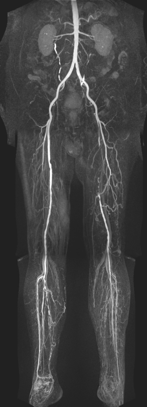
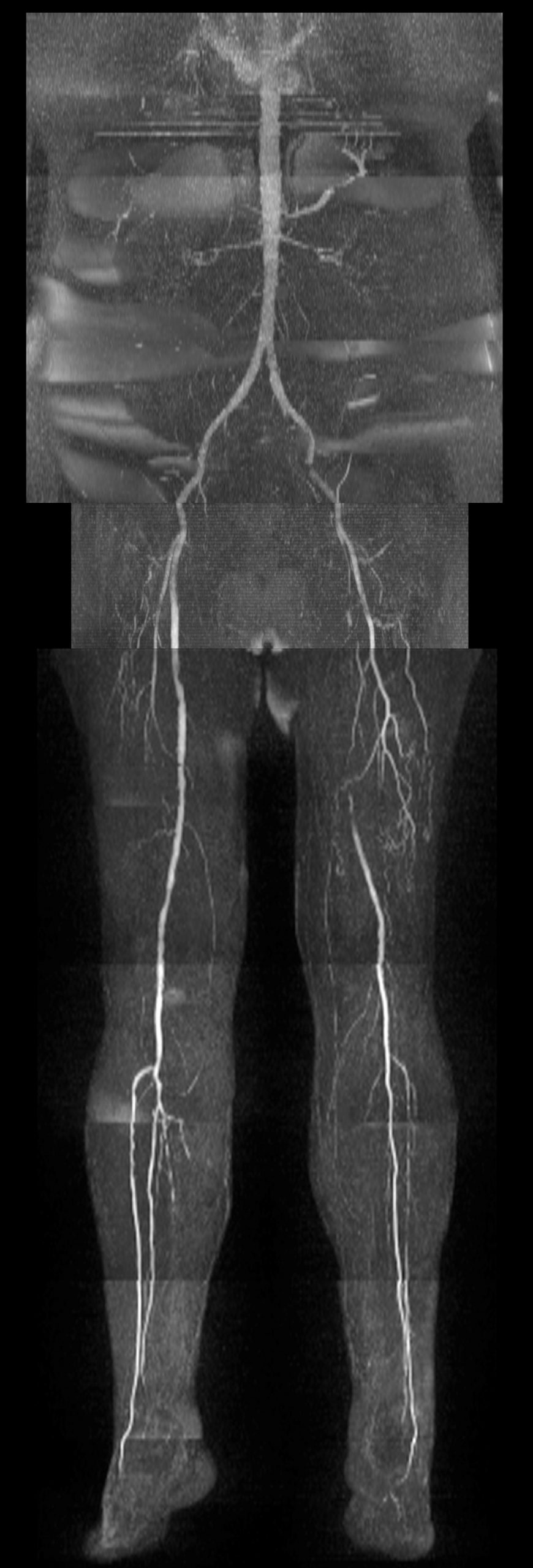
Left peripheral occlusive vascular disease with segmental occlusion of the superficial femoral artery and single vessel below knee run-off to the foot via the peroneal artery. The left posterior tibial artery is occluded while the anterior tibial artery demonstrates short segment occlusion in the proximal calf and reconstitution in the mid-calf with dorsalis pedis patency.
Right peripheral occlusive vascular disease is less severe with mild multifocal outflow disease involving the superficial femoral and popliteal arteries and two vessel run-off to the foot via a mildly diseased peroneal artery and a severely diseased anterior tibial artery with dorsalis pedis patency. The posterior tibial artery is severely diseased proximally and occludes in the mid-calf with incomplete distal reconstitution.
There is excellent agreement between non-contrast QISS MR angiography and contrast-enhanced MR angiography. Challenges with venous signal contamination are apparent in the calf station of the bolus-chase MR angiogram, with overlap that limits assessment of the posterior tibial arteries in particular. However, this is obviated by the use of initial time-resolved MR angiography which clearly separates out the arterial and venous phases of contrast enhancement. Non-contrast QISS MRA image quality was adequate for diagnosis but overall inferior to contrast-enhanced MR angiography, particularly in the assessment of the abdominal aorta and its branch vessels.
An additional point of interest is that image quality in this patient was excellent using a single dose of gadobutrol, diluted 1:1 with normal saline in this patient with moderate renal insufficiency.
Discussion:
Non-contrast QISS MRA is a robust technique for evaluation of lower extremity peripheral vascular disease. Image quality with current commercially available versions of QISS is dependent on a regular heart rhythm, as beat-to-beat variability can manifest as bilateral signal loss. In such situations, symmetry of such signal loss would be a clue that artifact rather than disease was present. Both non-contrast MRA and single dose contrast-enhanced MR angiography techniques are reliable, robust solutions to evaluate the severity and extent of lower extremity peripheral vascular disease.
Figures:
1- BCMRA – Bolus chase, 3 station contrast-enhanced MRA

2- QISS MRA – Non-contrast MRA with QISS technique

3- TRMRA – Time resolved MRA of calves with TWIST technique
